
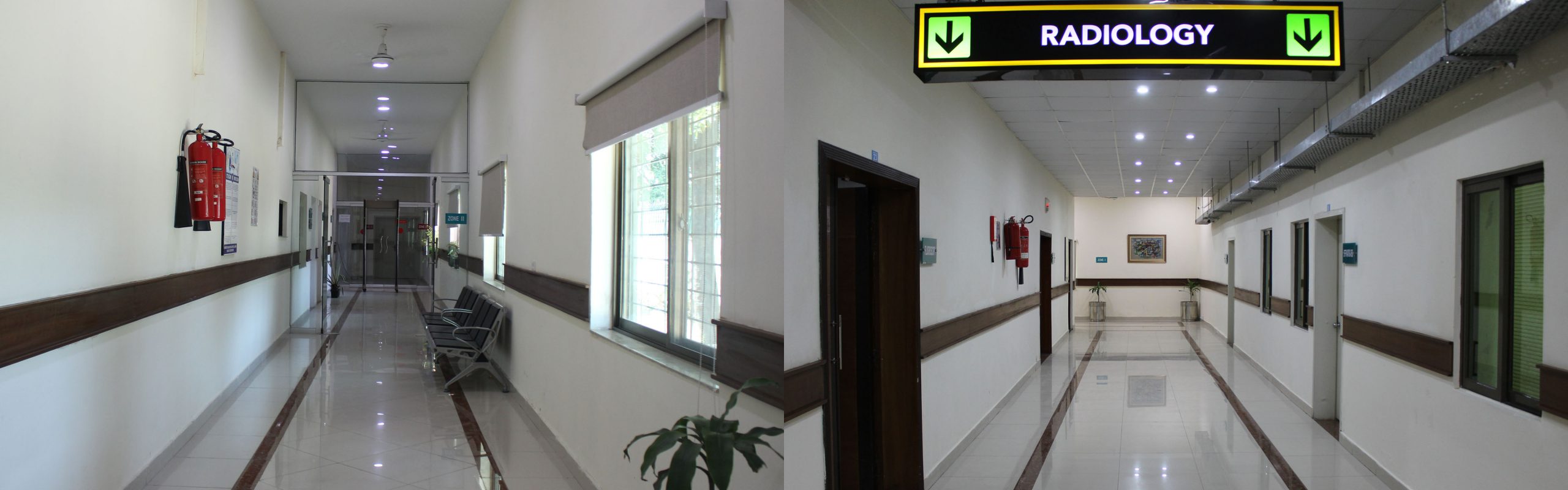
Department of Radiology and Diagnostic Imaging is fully equipped with state-of-the-art machines.
For wards and intensive care units, sophisticated mobile X-Ray machine are available. Gray scale and Doppler Ultrasound is also available along with TVS facility. 64 Slice latest CT scan is functioning in the department since Jan, 2018. Radiology Department is available for different medical specialties from General Practice, Orthopedics, Sports Medicine, Neurology, Internal Medicine, Gynaecology, Pediatrics, ENT, Pulmonology, Dental and Urology. We are specialized in Brain MRI, Spine MRI, Knee MRI, Full body MRI scan, Same day MRI scan, MRI breast, spine x ray, Ultrasound scan at our radiology clinic. Generally almost all kinds of cases from different doctors of different specialties.
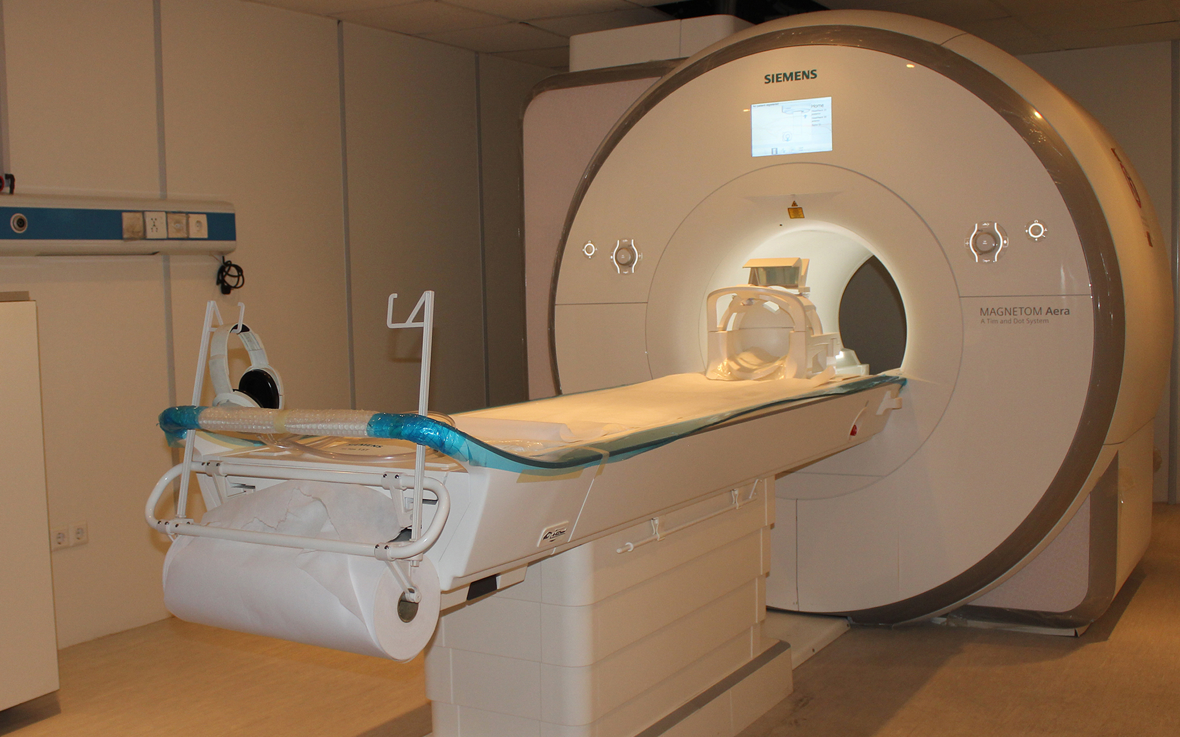
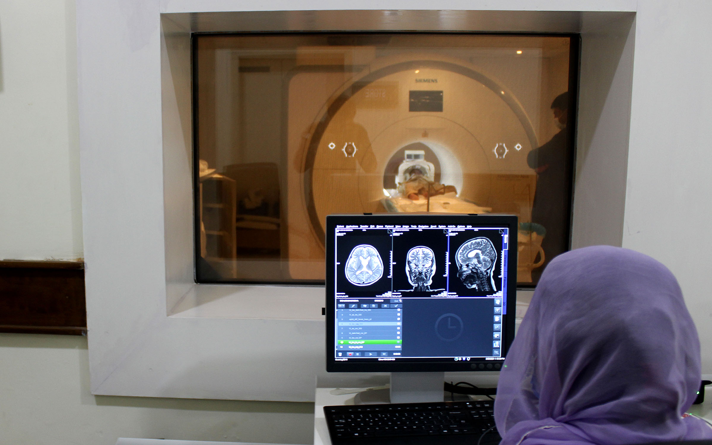
MRI
We are providing MRI scans and specialized studies which include Diffusion MRI, Fluid Attenuated Inversion Recovery (FLAIR), Magnetic resonance angiography, MRCP, MRI Liver, MRI abdomen, MRI prostate, MR spectroscopy, MRI IAMs, CSF cineflow study, MRI for brachial plexus, MRI breast, and MRI for trigeminal neuralgia and many more.
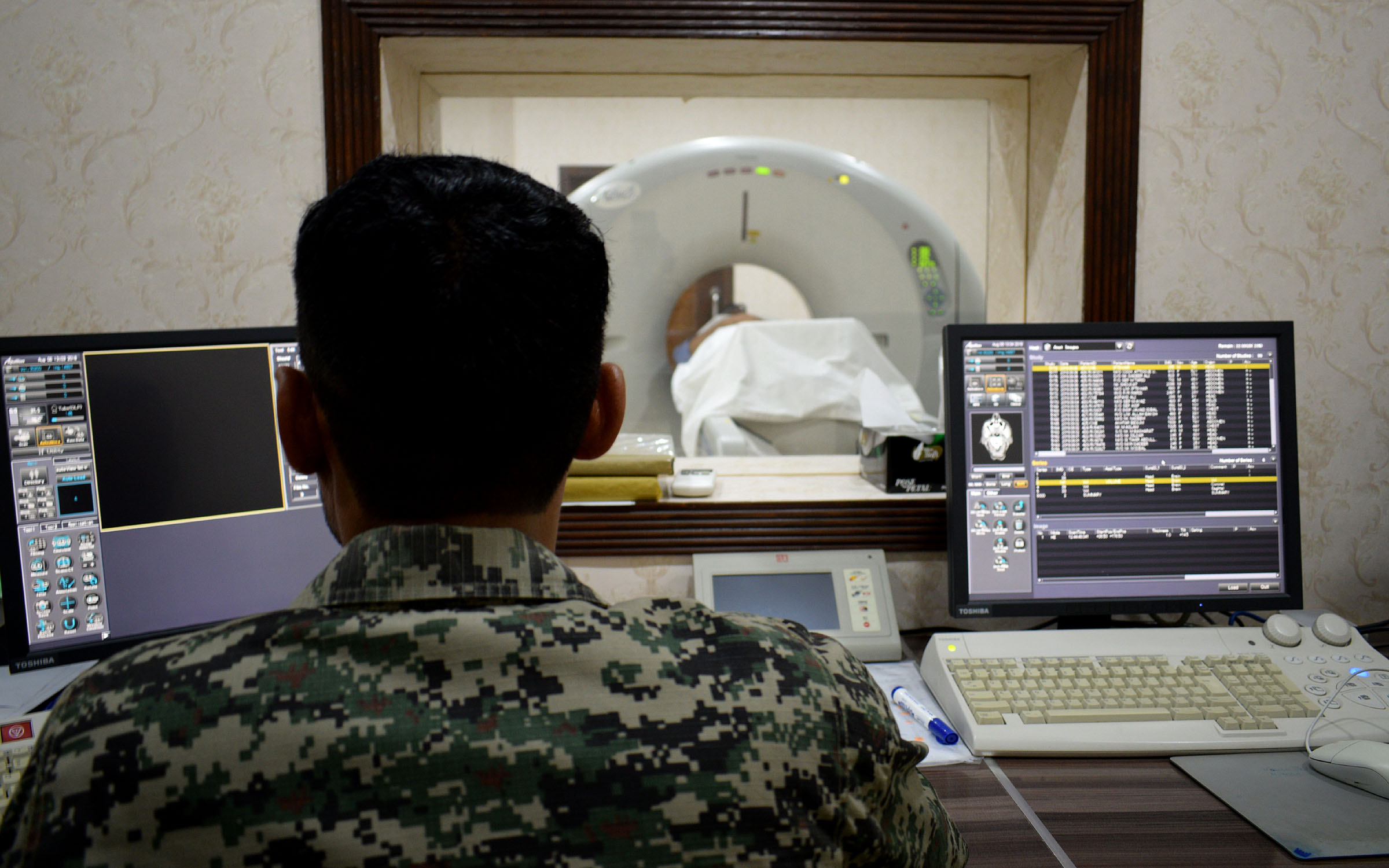
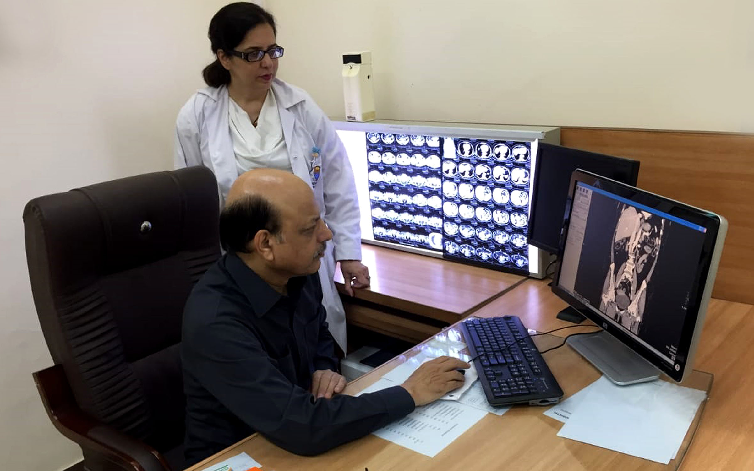
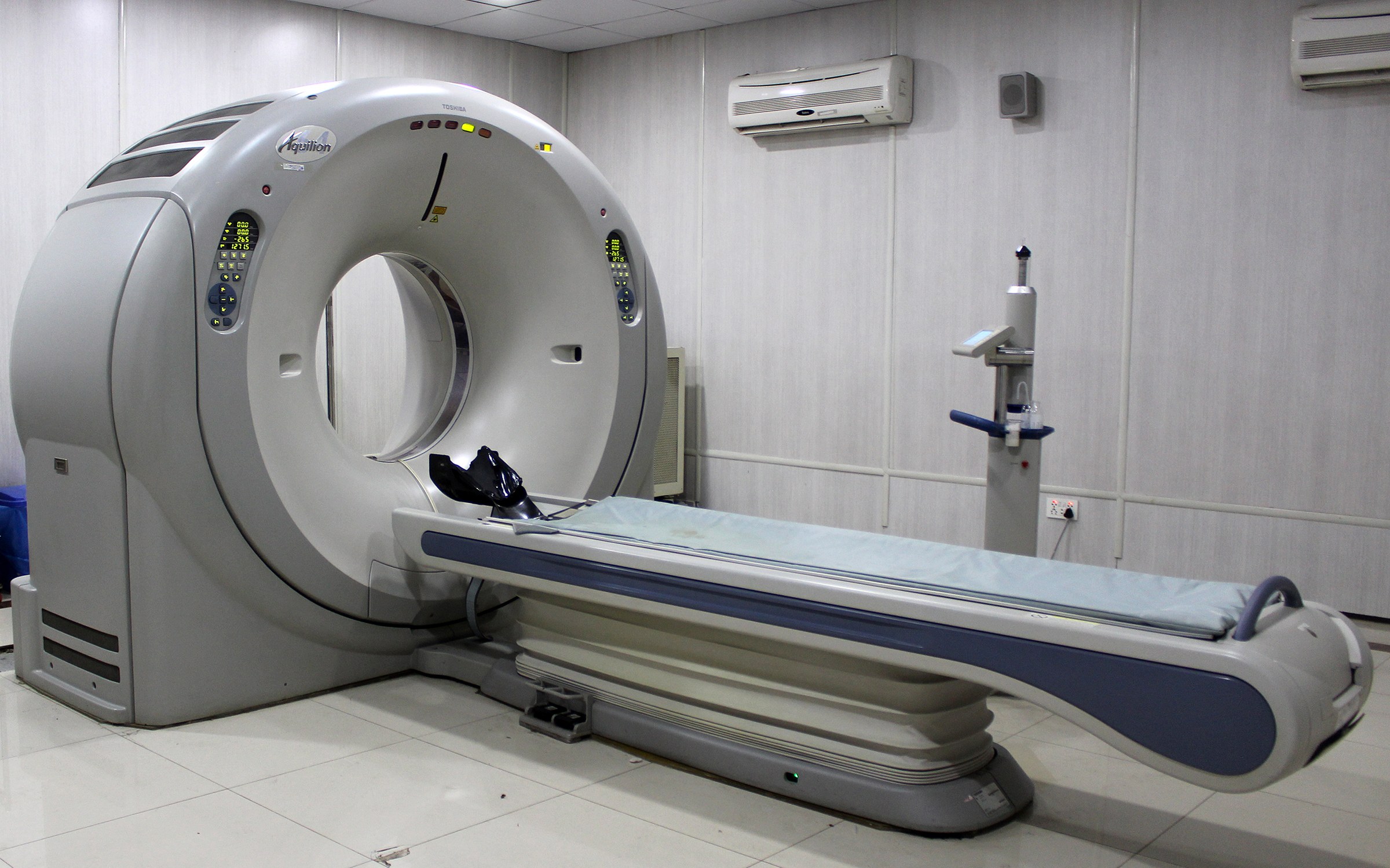
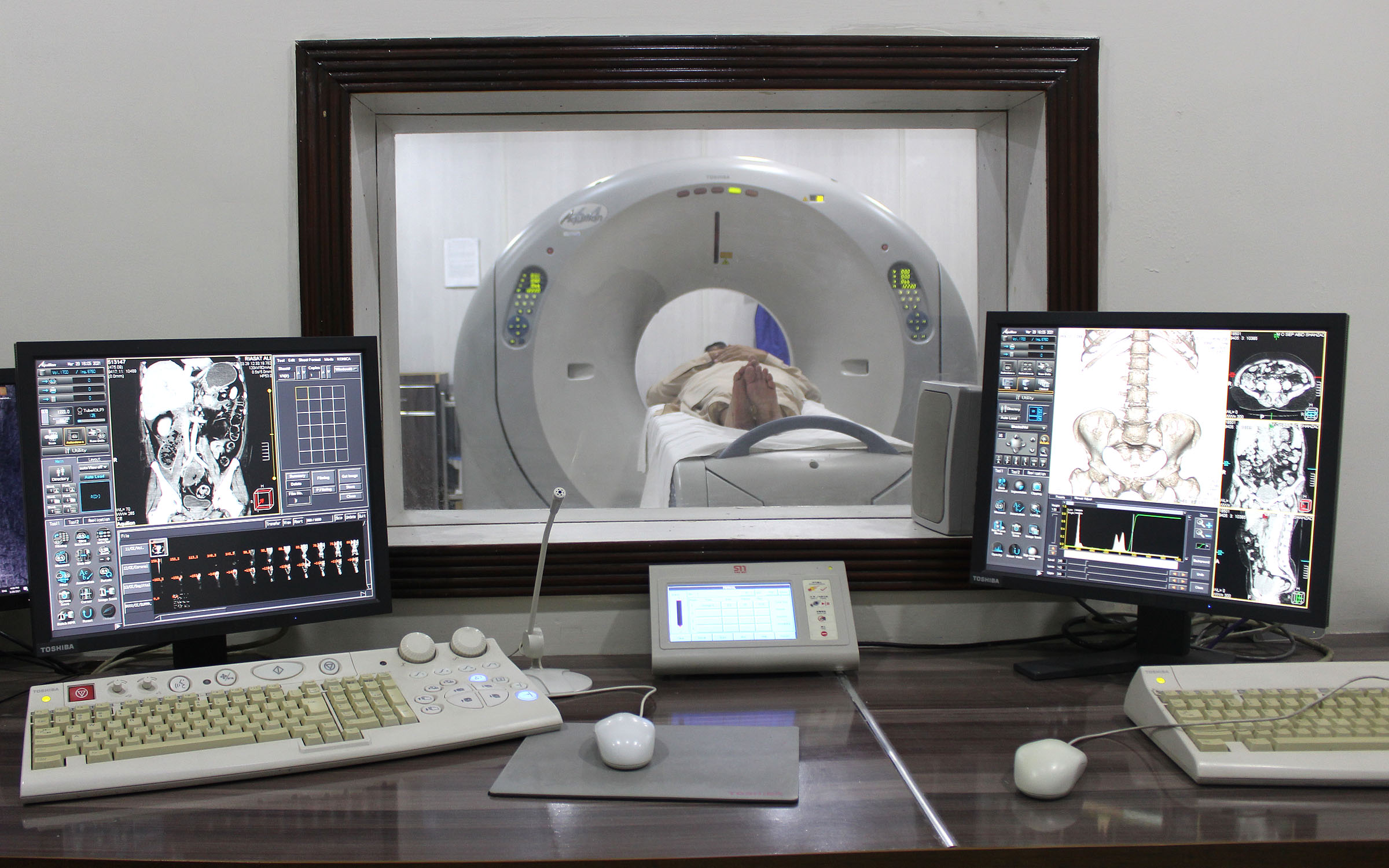
CT SCAN
Radiologists can more easily diagnose diseases such as cancers, cardiovascular diseases, infectious diseases, appendicitis, trauma and skeletal disorders via Computed Tomography (CT) scanner that provides uncompromised image quality and outstanding clinical performance.
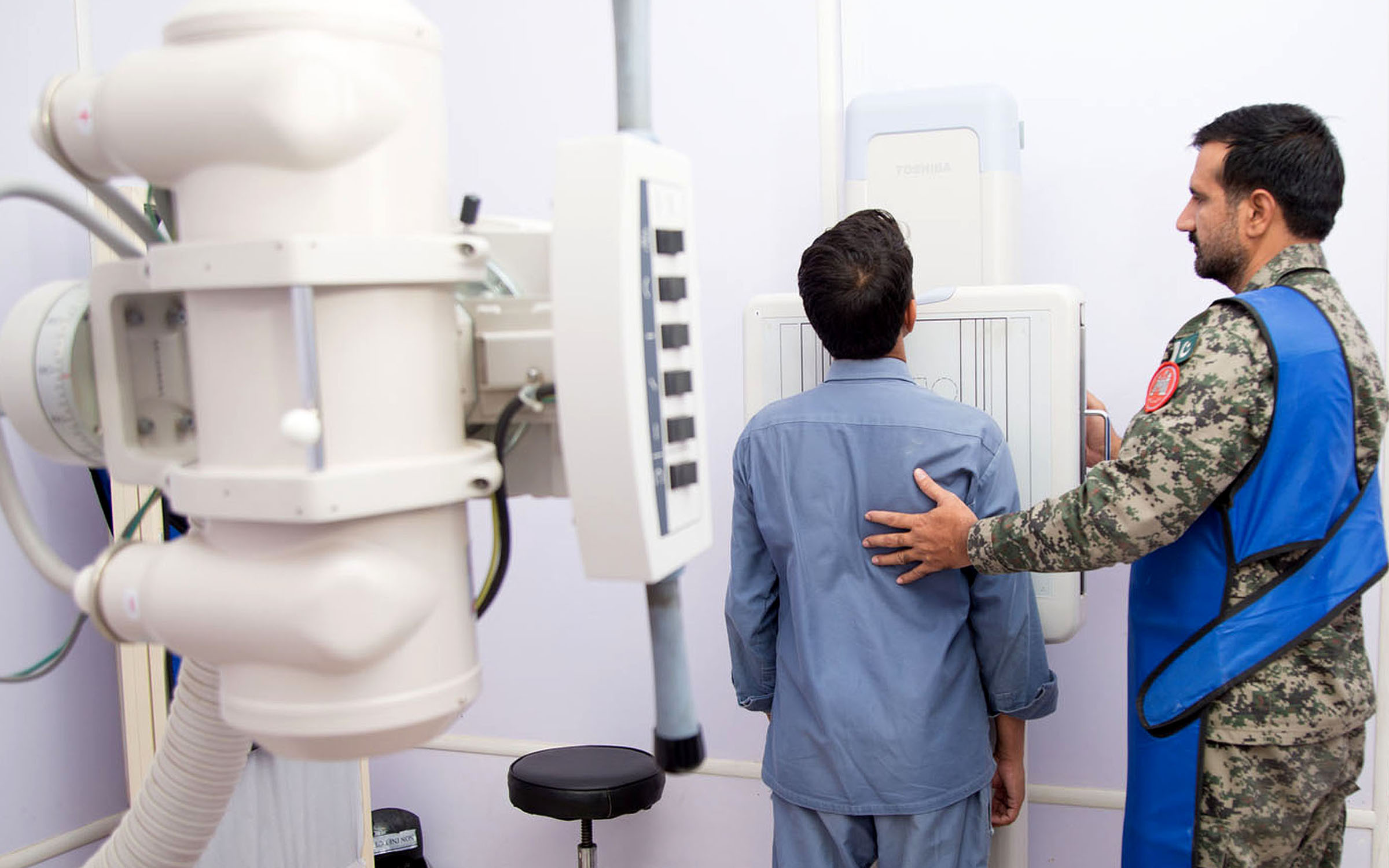
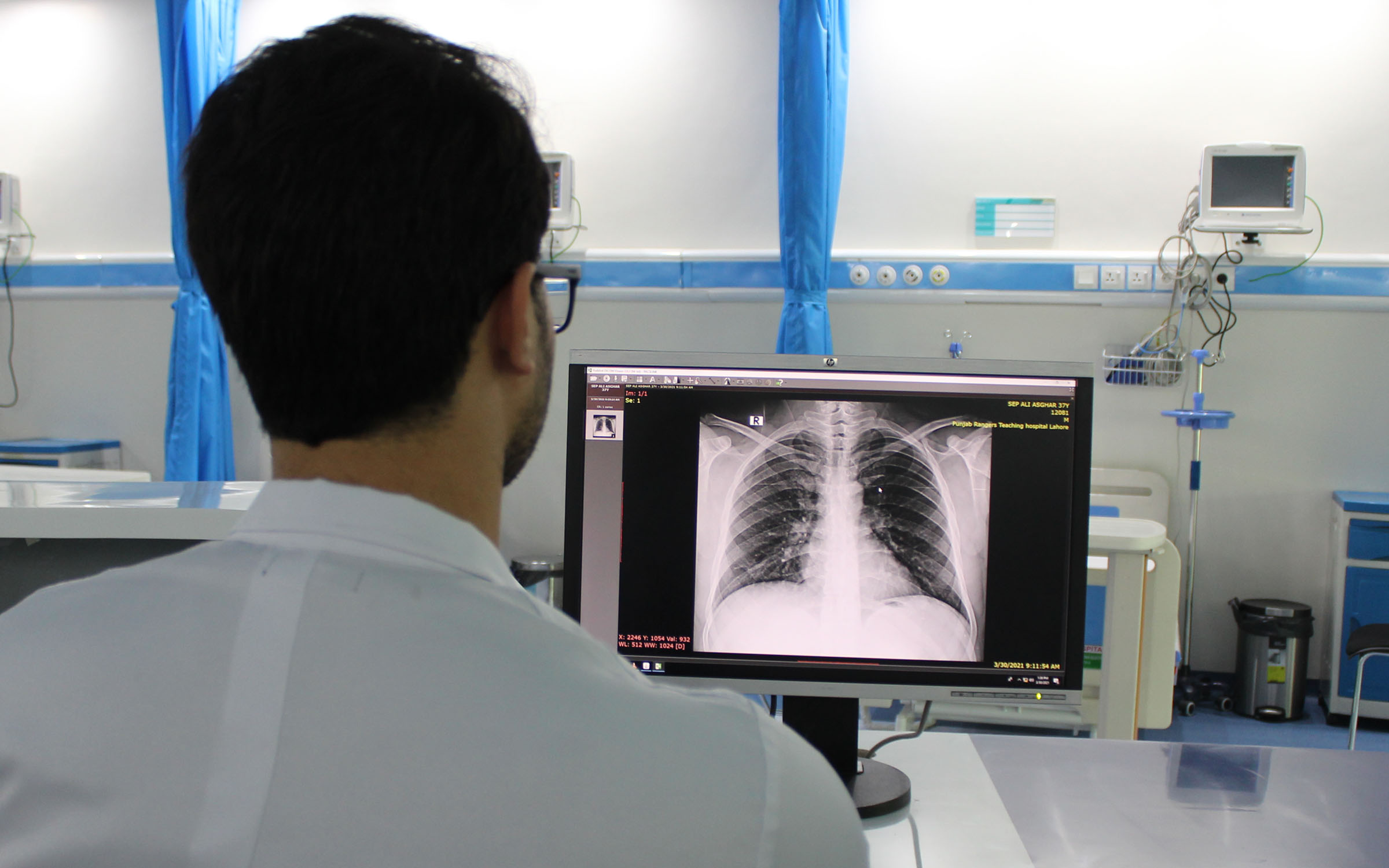
X – RAY
All routine X-rays such as bone and chest X-rays do not require appointments and walk-in service is provided round the clock. Special X-ray procedures like Fluoroscopy exams require a scheduled appointment including, Barium swallow, Barium meal, Barium enema, Hysterosalpinogram, IVP, skeletal survey, small bowel studies, MCUG, ureterograms, fistulograms and sonograms, etc
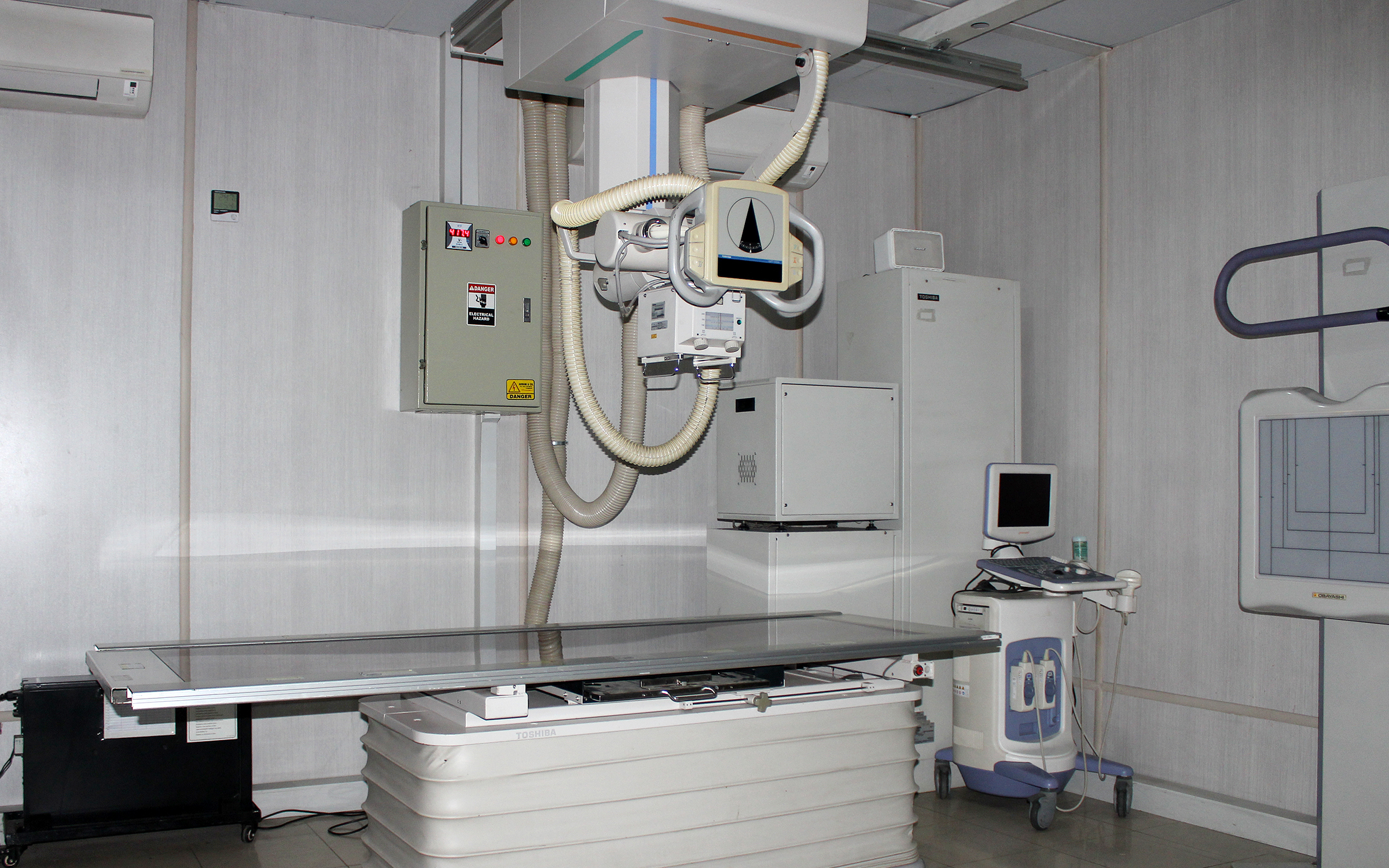
FLUOROSCOPY
Punjab Rangers Teaching Hospital offers Fluoroscopy service which is a digital imaging technique that takes real time (live) moving images of patients’ internal structures using X-rays (radiation). The fluoroscope is a flat table with a camera that pulls over the patient and creates a tunnel. The radiologist or technologist moves the camera up and down to best see the area being examined.
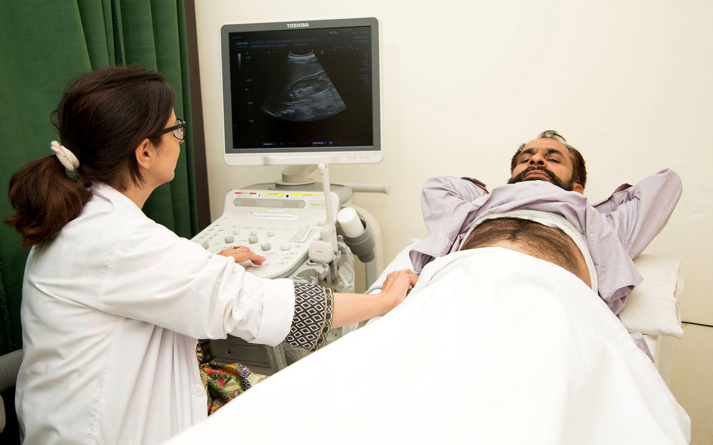
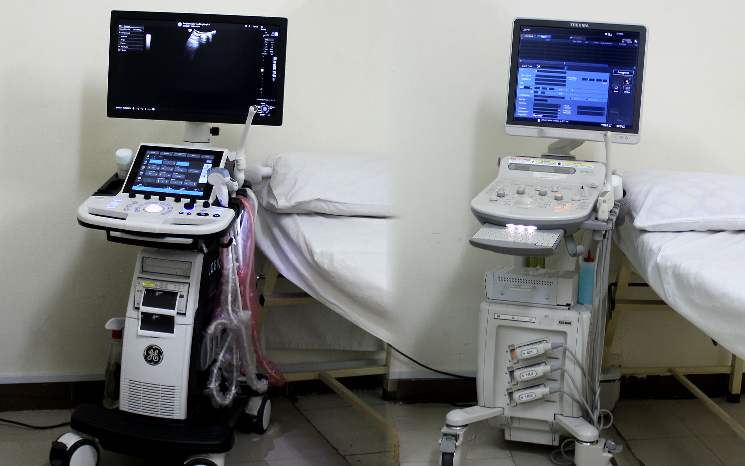
ULTRASONOGRAPHY
The Ultrasonography service at Punjab Rangers Teaching Hospital is equipped with four ultrasound and one US Doppler room. The ultrasound image is produced by sending sound waves of 1-10 million Hertz through a transducer by placing it over the structures of the body. The sound waves either get absorbed or get bounced back to crystals in the head of the transducer. For example, sound waves go through areas that are hollow or fluid-filled, such as the bladder and blood vessels. These areas appear black on the screen. Areas filled with tissue allow some penetration and refraction of sound and produce a grayish-white image. Hard structures, such as bone, produce a bright white image as the sound waves completely bounce back to the transducer.

In pregnancy, this allows providers to have an image of the woman’s womb. The amniotic fluid will appear black, which enhances the bones and tissues of the baby, which will appear white. The doctor can assess how well the baby is developing, it can determine the gender of the baby and detect any abnormalities. Along with the facility of ultrasonography for Obs and Gyne Patients, we also have services for Emergency patients who are provided with the services as soon as possible.
Real-time imaging is necessary to spot abnormalities and evaluate tissues and organ health during check-ups. Hence, a reliable and precise ultrasound is necessary for patients seeking medical advice and diagnosis.
Radiology Department offers a wide range of imaging diagnostic services, including top-notch ultrasound scan. As a leading diagnostic clinic, our mission is to provide accessible diagnostic and testing services to patients at price points that they can afford. Combining our expertise and our latest equipment, we can provide high-quality ultrasound imaging based on your need.
Our ultrasound service include 3D and 4D ultrasound and are used in different tests. Using sound waves, an ultrasound device can create images of internal organs and tissues in the body for abnormality detection. It is also used for fetal health tracking. We provide affordable and cheaper ultrasound services with best-in-class image quality with 3D and 4D functionality.
Radiology Investigation provided by the department are:
All kinds of digital plain radiography.
Contrast studies, like IVU and HSG.
Whole body CT scan, plain and with contrast.
Ultrasound for Abdomen, Chest, Pelvis, male and female reproductive organs.
Macular-skeletal ultrasound.
Doppler Studies for Limbs, Carotids and all others.
Ultrasound guided FNAC’s and drainage procedures like, pleural effusion taping, ascetic fluid taping, percutaneous nephrostomies and abscess drainage.
Routine Procedures.
Upper Abdomen (liver, gallbladder, pancreas, spleen, biliary tree, kidney, adrenal glands, abdominal aorta).
Pelvic (uterus, ovaries, urinary bladder, prostate gland).
Transvaginal Sonography.
Transrectal Sonography.
Thyroid Gland.
Classification of Chest Densities (pleural effusion).
Breast Sonography.
Transcranial Sonography for infants.
Soft tissue or small parts sonography.
Scrotal Ultrasound.
Musculoskeletal Ultrasound.
Special Procedures.
Doppler Sonography.
Extremities.
Faculty Members
Maj (R) Dr Asma Tariq
Associate Professor
Dr. Abdul Qayyum
Assistant Professor
Dr. Tarab Kamran
Senior Registrar